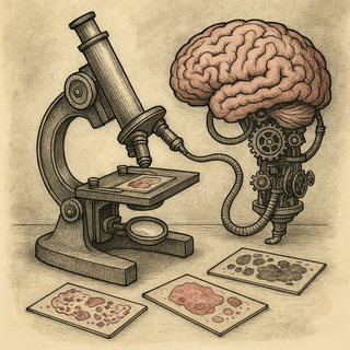
A pathologist looks at a tissue slide under a microscope, evaluates cell morphology, architecture, staining patterns, and invasion depth, then renders a diagnosis. This process has been the backbone of cancer diagnosis for over a century. It is also subjective, time-consuming, and limited by human fatigue and inter-observer variability. So naturally, someone trained an AI to do it. And it turns out the AI has some opinions.
Digital Pathology 101
Before AI can read a slide, the slide has to become digital. Whole-slide imaging (WSI) scanners capture tissue sections at 20x or 40x magnification, producing images that are absolutely enormous - a single slide can generate a file of 2-5 gigabytes containing billions of pixels. This is not your phone camera's resolution; it is closer to satellite imagery of a small city, except the city is made of cells and some of them are trying to kill you.
The digitization step alone was controversial for years. Pathologists were skeptical. Regulatory bodies were cautious. But FDA clearances for digital pathology systems starting in 2017 opened the floodgates, and the field has not looked back.
What AI Actually Does With a Slide
The most straightforward application is automated cancer detection. Deep learning models - typically convolutional neural networks and increasingly vision transformers - are trained on thousands of annotated slides to recognize malignant tissue. For breast cancer metastasis detection in lymph nodes, prostate cancer grading (Gleason scoring), and cervical cancer screening, AI systems now perform at or above the level of board-certified pathologists in controlled studies.
But detection is just the beginning. AI can extract information from slides that human eyes cannot reliably perceive.
Mutation prediction from morphology. Multiple groups have shown that deep learning models can predict specific genetic mutations - BRAF, TP53, microsatellite instability, EGFR, and others - directly from H&E-stained tissue images. The model is finding subtle morphological patterns associated with these mutations that pathologists were never trained to see. This could reduce the need for molecular testing in some cases or at least prioritize which patients need it.
Prognosis prediction. AI models can predict patient survival from tissue morphology alone, sometimes outperforming traditional clinical staging. The models learn patterns in the spatial arrangement of cells, the tumor-stromal interface, and immune cell infiltration that correlate with outcomes. In colorectal cancer, breast cancer, and lung cancer, AI-derived prognostic scores are being validated in large cohorts.
Treatment response prediction. Perhaps the most valuable application: predicting which patients will respond to specific therapies. Models trained to predict immunotherapy response from pre-treatment biopsies are in development, analyzing immune cell infiltration patterns and tumor architecture to estimate the likelihood of benefit from checkpoint inhibitors.
The Foundation Model Wave
The latest development is pathology-specific foundation models - large AI models pre-trained on millions of pathology images that can then be fine-tuned for specific tasks. CONCH, UNI, Virchow, Prov-GigaPath, and similar models have been trained on datasets of 100,000+ slides and can perform diverse downstream tasks with minimal additional training. This is the same paradigm that gave us GPT for language now applied to tissue images.
These foundation models are particularly good at tasks with limited labeled data. Instead of needing 10,000 annotated slides for each new cancer subtype, you can fine-tune a foundation model with a few hundred examples. For rare cancers where annotated data is scarce, this is a significant advantage.
The Reality Check
AI is not replacing pathologists. Anyone who tells you otherwise is selling something (probably AI software). What it is doing is augmenting them - flagging suspicious regions, prioritizing worklists, providing quantitative measurements, and offering second opinions on difficult cases.
The practical challenges are real. Models trained at one institution often perform poorly at another due to variability in staining and scanning equipment. Clinical validation lags behind technical publications. And reimbursement - who pays for AI analysis and how much - remains unsolved. These are not technical questions, but they determine whether the technology actually gets used.
The Data Pipeline Challenge
The sheer volume of imaging data generated by digital pathology is staggering. A busy pathology department digitizing all its slides produces terabytes per week. Storing, managing, annotating, and analyzing that data requires infrastructure most hospitals do not currently have. If you are handling the non-image side of pathology research - papers, reports, protocols - tools like pdfb2.io can at least tame the document pile while you figure out your petabyte storage strategy.
The trajectory is clear: within a decade, AI-assisted pathology will be standard in well-resourced centers. The interesting question is whether it reaches the places that need it most - hospitals without enough pathologists and clinics where expertise is scarce. That is where the real impact lives.
References
- Lu MY, Chen B, Williamson DFK, et al. A visual-language foundation model for computational pathology. Nat Med. 2024;30(3):863-874. DOI: 10.1038/s41591-024-02856-4 | PMID: 38504017
- Coudray N, Ocampo PS, Sakellaropoulos T, et al. Classification and mutation prediction from non-small cell lung cancer histopathology images using deep learning. Nat Med. 2018;24(10):1559-1567. DOI: 10.1038/s41591-018-0177-5 | PMID: 30224757
Disclaimer: This blog post is for informational and educational purposes only. It is not medical advice. Always consult a qualified healthcare professional for clinical decisions.
Get cancer research delivered to your inbox
The best new studies, explained without the jargon. One email per week.